


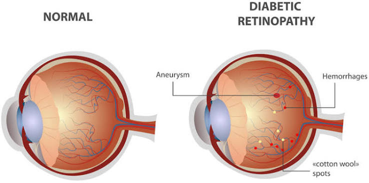
Diabetic Retinopathy is one of the leading causes of preventable blindness in working-age adults. Prolonged elevation of blood glucose levels weakens the tiny blood vessels supplying the light-sensitive retina at the back of the eye.
Understanding the Stages of Retinal Damage
Diabetic retinopathy generally progresses through two main phases:
- Non-Proliferative Diabetic Retinopathy (NPDR): Early stage where retinal blood vessels swell, leak fluid or blood, and form microaneurysms. Fluid accumulation in the center of vision (Diabetic Macular Edema - DME) leads to blurred vision.
- Proliferative Diabetic Retinopathy (PDR): Advanced stage where blocked blood vessels trigger the growth of fragile, abnormal new blood vessels. These can bleed into the vitreous gel (Vitreous Hemorrhage) or cause tractional retinal detachment.
Silent Danger: Why Annual Exams are Essential
In its early stages, diabetic retinopathy causes zero pain and often no visible change in vision. Significant damage can accumulate silently before you notice any blurriness. That is why every diabetic patient must undergo an annual comprehensive dilated eye examination.
Advanced Retina Diagnostics at EyeNexa
- High-Resolution Digital Fundus Photography: Captures detailed wide-field images of the retina to track micro-vascular changes over time.
- OCT Macular Mapping: High-speed 3D optical coherence tomography measuring macular thickness in microns to detect subtle edema.
- Fundus Fluorescein Angiography (FFA): Specialized imaging using dye to pinpoint exact leakage sites and vessel blockages.
"Strict blood sugar control combined with annual dilated retinal screenings can reduce the risk of severe vision loss from diabetic retinopathy by over 90%." — Dr. Kumar Sambhav
Cutting-Edge Retinal Treatments
- Anti-VEGF Intravitreal Injections: Targeted anti-VEGF medications (e.g. Ranibizumab, Aflibercept) injected painlessly to stop vessel leakage and shrink abnormal vessels.
- Targeted Laser Photocoagulation: Precision laser treatment to seal leaking blood vessels or shrink abnormal new vessels in PDR.
- Micro-Incision Vitrectomy Surgery (MIVS): Advanced surgical clearance of persistent vitreous blood or scar tissue.